
How Palm Harbor Dentists Handle All-on-4 Cases With Existing Gum Disease: Clinical Strategies and Patient Care
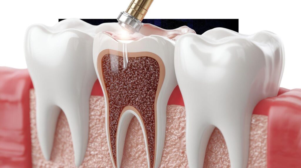
If you have gum disease but want all on 4 dental implants in Palm Harbor, FL, dentists don’t automatically rule you out. They evaluate, treat, and sequence care so implants can succeed.
Your dental team will stabilize periodontal disease first, then assess bone, infection risk, and overall health. This determines whether immediate All-on-4, delayed placement, or alternative solutions best protect your long-term outcome.
You will learn how clinicians grade gum disease, when they recommend scaling, antibiotics, or surgery before implant work. Specialists collaborate to plan implant position and bone grafting when needed.
Expect clear guidance on what makes you a suitable candidate and what compromises might exist. You will also learn about follow-up and maintenance support to keep your new arch healthy.
Identifying Types and Severity of Gum Disease
You need to know which form of gum disease you have and how deep the infection extends. Local or systemic factors that increase your risk are also considered.
Those specifics determine whether All-on-4 planning proceeds now, after treatment, or with modified protocols.
Clinical Assessment Techniques
Your dentist will perform a full periodontal charting to measure pocket depths at six points per tooth. Probing depths of 3 mm or less usually indicate healthy tissue; depths ≥4 mm suggest periodontitis and require intervention.
Bleeding on probing and the presence of suppuration (pus) signal active inflammation and infection that must be controlled before implant placement.
Visual inspection checks for gingival recession, swelling, and fistulae. Tooth mobility is graded (Class I–III) to assess loss of attachment and occlusal forces that could affect implant planning.
Clinical attachment loss and furcation involvement are documented because they change prognosis. These may necessitate regenerative procedures or extractions prior to All-on-4.
Diagnostic Technologies
Radiographs provide essential information about bone levels and architecture. Periapical and bitewing X-rays show vertical and horizontal bone loss.
A full-mouth series or CBCT offers three-dimensional views critical for implant site assessment. CBCT helps you and your clinician evaluate ridge width, sinus position, and residual bone quantity for All-on-4 angulation and implant length decisions.
Microbiological testing and inflammatory biomarkers can be used selectively to identify aggressive pathogens or persistent inflammation. Photographic records and periodontal software track progression over time, guiding decisions about timing of implant placement and the need for adjunctive therapies.
Common Risk Factors
Smoking increases pocket depth and bone loss and reduces healing capacity after implant surgery. If you smoke, your clinician will discuss cessation or delayed treatment to improve outcomes.
Poor oral hygiene and irregular professional cleanings allow biofilm to mature. You must demonstrate effective plaque control before moving forward with All-on-4.
Systemic conditions like uncontrolled diabetes impair immune response and bone healing. Your provider will require medical clearance and glucose control targets prior to implant surgery.
Medications such as bisphosphonates, anticoagulants, or immunosuppressants also alter risk. These may change surgical timing or require collaboration with your physician.
Candidacy and Evaluation for All-on-4
You will need a thorough clinical and radiographic evaluation to determine if All-on-4 suits you. The process focuses on your periodontal health, jawbone quantity and quality, and overall medical status.
Criteria for Patient Suitability
You must be able to tolerate minor oral surgery and follow postoperative care. Typical favorable factors include adequate general health (controlled diabetes, no recent head/neck radiation), commitment to oral hygiene and regular dental visits, and sufficient opposing dentition or willingness to restore both arches if needed.
Absolute and relative contraindications matter. Active, uncontrolled systemic disease or heavy smoking can reduce success rates.
Previous failed implants or extreme parafunctional habits (severe bruxism) may require adjunctive measures such as occlusal guards, expanded implant number, or alternative prosthetic designs.
Impact of Active Periodontal Disease
Active periodontal infection increases the risk of implant failure and prosthesis complications. You must have periodontal treatment before implant placement—scaling/root planing, localized surgery, and sustained maintenance.
Dentists will measure pocket depths, bleeding on probing, and presence of suppuration. Persistent deep pockets (>6 mm) or wide bacterial reservoirs typically delay All-on-4 until infection control is achieved.
In some cases, staged treatment places implants only after stable periodontal parameters persist across multiple maintenance visits.
Evaluating Bone Density and Structure
Your jawbone must provide primary stability for the four implants. Assessment uses CBCT and periapical radiographs.
Clinicians evaluate bone height and width at planned implant sites, cortical thickness and trabecular density, and sinus position and inferior alveolar nerve course.
If bone quantity or quality is insufficient, options include bone grafting, zygomatic implants, angled implant placement, or using longer/tilted implants as in the All-on-4 protocol. The final plan balances achieving at least 30–35 Ncm insertion torque with minimizing additional surgical complexity for you.
Pre-Treatment Solutions for Gum Health
You will see targeted options that reduce infection, rebuild damaged tissue when needed, and control bacterial load before implant placement. Each approach aims to lower pocket depths, arrest bone loss, and create a stable soft-tissue environment for All-on-4 prosthetics.
Non-Surgical Periodontal Therapies
Non-surgical therapy focuses on mechanical removal of plaque and calculus to reduce pocket depth and inflammation. Your dentist or hygienist will perform scaling and root planing (SRP) under local anesthesia, often over one to three visits depending on disease extent.
Adjunctive tools may include ultrasonic scalers, air polishing, and localized delivery of antimicrobials placed into pockets. You will receive customized oral hygiene instruction to maintain gains.
Clinics often re-evaluate 4–8 weeks after SRP to measure probing depths and bleeding on probing. If pockets remain ≥5 mm with bleeding, your team will discuss surgical options to reach the stability required for All-on-4.
Surgical Interventions
Surgical therapy addresses persistent deep pockets, vertical bone defects, and insufficient keratinized tissue that non-surgical care cannot fix. Your periodontist may perform flap surgery (pocket reduction) to gain access for debridement and reshape bone contours.
For bone defects that threaten implant support, they may use guided bone regeneration with bone grafts and barrier membranes. Soft-tissue grafting improves the width of keratinized tissue around future implants to enhance hygiene access and long-term stability.
Surgeries typically require pre-op imaging and local or conscious sedation. Healing times vary: soft-tissue grafts 2–4 weeks for initial closure, bone graft maturation often 4–6 months before implant placement or immediate loading decisions.
Antibiotic Regimens
Antibiotics support mechanical and surgical therapies but do not replace them. Your provider will choose systemic or local agents based on microbial profile, disease severity, and medical history.
Common systemic protocols include a short course of amoxicillin plus metronidazole for aggressive or refractory cases, or doxycycline where beta-lactam allergy exists. Localized antibiotics such as minocycline microspheres or doxycycline gel can be placed directly into residual pockets.
You must follow dosing exactly and disclose medications, allergies, and conditions like diabetes. Practitioners will reassess clinical response within weeks and avoid unnecessary prolonged antibiotic use to reduce resistance risk.
Planning the All-on-4 Procedure With Existing Gum Concerns
You will need a targeted plan that controls infection, preserves or augments bone, and delivers a prosthesis tailored to your mouth. The team will coordinate sequencing, prosthetic design, and infection control to reduce failure risk and speed recovery.
Treatment Sequencing Strategies
Your dentist will stage care based on infection severity and bone condition. If active periodontitis exists, expect initial periodontal therapy: scaling and root planing, localized antibiotic delivery, and at-home antimicrobial rinses until inflammation and bleeding subside.
Re-evaluation typically follows 4–8 weeks to confirm tissue stability. When bone loss or tooth mobility persists, the clinician may extract hopeless teeth and place implants either immediately with careful debridement or after a healing interval.
Immediate placement suits controlled infection and adequate bone. Delayed placement (3–6 months) suits extensive infection or need for bone grafting.
Your surgeon will also decide if simultaneous bone grafting or sinus lift is safe based on residual bone height and infection control.
Customized Prosthetic Design
Your prosthesis will reflect surgical choices and oral conditions. The lab and clinician will design a fixed full-arch prosthesis that compensates for implant angulation, soft-tissue contours, and any grafted areas.
Material selection (acrylic over titanium bar, monolithic zirconia, hybrid options) balances strength, hygiene access, and cost. Occlusal scheme gets deliberate planning to reduce overload on the four implants.
Your dentist will specify a prosthetic emergence profile and hygiene access points to permit effective cleaning under the bridge. Temporary prostheses used during healing will protect implants while allowing you to test function and aesthetics before final fabrication.
Mitigating Infection Risk
You will follow a strict infection-control protocol before, during, and after surgery. Preoperative measures may include systemic antibiotics for high-risk cases, chlorhexidine rinses, and targeted periodontal treatment to lower bacterial load.
Intraoperative steps include thorough socket debridement, sterile technique, and possibly use of antibiotic-impregnated graft materials. Postoperative management emphasizes wound stability and plaque control.
Your follow-up schedule will be more frequent—weeks 1, 2, and monthly until osseointegration—so clinicians can catch early signs of peri-implantitis. Long-term, you must commit to professional maintenance every 3–6 months and meticulous home care to protect your implants from recurrent infection.
Interdisciplinary Collaboration in Complex Cases
You’ll rely on coordinated care between specialists to manage infection, preserve bone, and deliver a stable All-on-4 prosthesis. Clear roles, shared diagnostics, and staged treatment timing reduce risk and improve long-term success.
Coordinating With Periodontists
You and the periodontist must assess periodontal stability before implant placement. That starts with a full-mouth periodontal chart, radiographic bone-level analysis, and microbial risk assessment when indicated.
Agree on infection-control criteria—pocket depths, bleeding on probing, and radiographic bone loss thresholds—so you know when to proceed or delay. Plan treatment sequencing together.
Options include staged periodontal therapy followed by implant placement, simultaneous extractions with guided bone regeneration, or extraction-socket preservation when needed. Decide who manages grafting, soft-tissue augmentation, and antibiotic protocols.
Document responsibilities in the patient chart. Specify follow-up intervals, maintenance frequency, and targeted clinical metrics.
Use shared imaging and digital treatment plans to align surgical guides and prosthetic emergence profiles.
Role of Dental Hygienists
Your hygienist is the clinical backbone for infection control and long-term maintenance. They perform initial nonsurgical therapy—scaling, root planing, and localized antimicrobial delivery—then monitor healing at short intervals.
Hygienists provide patient education on oral hygiene techniques customized for All-on-4 prostheses. They also teach self-exam signs for peri-implant inflammation.
Integrate hygienists into recall planning. Set specific maintenance protocols and have hygienists flag any tissue changes or radiographic bone loss for earlier specialist review.
Prosthodontic Considerations
You must coordinate prosthesis design with surgical planning to protect compromised tissues. The prosthodontist defines occlusion, cantilever length, and emergence profile to minimize biomechanical overload and facilitate hygiene access.
Choose provisional protocols that allow soft-tissue maturation and prosthetic contours that support keratinized tissue. Decide whether to use a fixed provisional on the day of surgery or a delayed prosthesis based on stability and infection control.
Specify material choices and retrievability. Use designs that allow easy removal for peri-implant treatment and repairs.
Document occlusal schemes, opposing dentition management, and contingency plans for future complications so all team members follow the same prosthetic objectives.
Patient Education and Postoperative Care
You will receive precise home-care steps, warning signs to watch for, and a clear schedule for clinical checks. These instructions focus on protecting implants, controlling gum disease, and catching complications early.
Oral Hygiene Instructions
You must avoid brushing the surgical sites vigorously for the first 48–72 hours. Use a soft-bristled brush and a non-abrasive toothpaste once healing begins.
Brush around abutments gently at a 45° angle to remove plaque without disturbing sutures or newly placed implants. Rinse with an antimicrobial mouthwash (chlorhexidine 0.12% if prescribed) twice daily for up to two weeks.
Use saline rinses after meals during the initial healing phase to keep debris clear. Interproximal cleaning is critical.
Use floss, interdental brushes (0.6–1.2 mm depending on space), or water irrigation devices as instructed to clean under the prosthesis and around implant collars. Replace or sterilize any hygiene aids after 7–10 days to prevent reintroducing bacteria.
Follow any dietary restrictions exactly: soft, protein-rich foods for the first week. Avoid hard, sticky, or crunchy items for at least 6–8 weeks.
Do not use tobacco or alcohol during early healing because both impede soft-tissue attachment and raise infection risk.
Monitoring for Complications
Watch for increased pain, swelling, persistent bleeding, fever, or a foul taste; these can indicate infection or implant failure and require immediate contact with your dentist. Mild soreness and transient swelling are normal; escalating symptoms after 48–72 hours are not.
Look for changes at the gum margin—redness, pus, or separation of the tissue from the implant should prompt an urgent visit.
Your clinician will teach you how to check mobility of the prosthesis and implants. Do not attempt to force or adjust the prosthesis yourself.
If you notice loosening, schedule an appointment the same day to avoid overload and bone loss.
Follow-Up Scheduling
Expect an initial post-op check within 24–72 hours to assess bleeding, suture integrity, and pain control. A 1–2 week visit will usually include suture removal and soft-tissue evaluation.
Your provider will schedule an implant stability assessment at 3 months (or earlier if guided by intraoral findings). The definitive prosthesis is finalized once osseointegration and periodontal stability are confirmed.
After the first year, plan maintenance visits every 3–4 months if you had prior gum disease. These visits include professional cleaning around implants, radiographic checks as indicated, and reinforcement of home-care techniques.
Long-Term Success and Maintenance Strategies
You will need a formal, regular professional maintenance plan plus a strict daily home-care routine to protect implants and surrounding gum tissue. Both focus on preventing peri-implantitis, monitoring bone levels, and maintaining prosthetic fit.
Routine Professional Cleanings
Your dentist or hygienist will schedule recalls every 3–6 months depending on your gum disease status and risk factors. During visits they will remove hard and soft deposits around implants using titanium or plastic instruments and ultrasonic tips designed for implant surfaces.
Expect periodic radiographs and probing to track marginal bone levels and detect early inflammation. Clinicians may perform localized antiseptic therapy (chlorhexidine rinse or gels) and adjust occlusion if prosthetic wear or bite changes threaten implants.
If signs of active peri-implant disease appear, your provider may increase visit frequency, prescribe antibiotics selectively, or refer you for surgical debridement and regenerative treatment. Early intervention prevents larger procedures later.
Guidelines for At-Home Care
Brush your prosthesis and remaining teeth twice daily with a soft-bristled brush and non-abrasive toothpaste to protect implant surfaces. Use an interdental brush (0.6–1.0 mm) or soft floss specifically made for implants to clean under and between the prosthetic framework.
Rinse once daily with an antimicrobial mouthwash if recommended by your clinician. Avoid long-term reliance on antiseptics without professional guidance.
Inspect your gums and prosthesis weekly for redness, swelling, persistent bleeding, or looseness and report changes promptly.
Control risk factors: stop smoking, manage diabetes, and maintain a balanced diet low in refined sugars. These steps reduce inflammation and support bone health around implants, improving the longevity of your All-on-4 restoration.
Emerging Technologies and Future Trends
You will see growing use of AI-driven diagnostics to assess peri-implant and periodontal risk before and after All-on-4 placement. These tools analyze imaging and chart data to identify inflammation patterns and predict implant failure risk.
Digital planning and guided surgery continue to improve precision. Cone-beam CT combined with surgical guides reduces placement error and lets you plan implant positions that avoid compromised bone while accommodating future soft-tissue management.
Minimally invasive antimicrobial therapies are becoming more common as adjuncts to debridement. Localized drug delivery systems, photodynamic therapy, and antiseptic-releasing biomaterials offer targeted control of pathogens without broad systemic exposure.
You will benefit from advances in regenerative materials for bone and soft tissue. Bioactive grafts, growth-factor–enhanced scaffolds, and collagen substitutes help rebuild support around compromised sites.
Remote monitoring and connected care shift postoperative follow-up into digital workflows. Teledentistry, wearable oral sensors, and patient apps let you track healing and reinforce hygiene.
Key practical considerations:
- Risk stratification first: Use new diagnostics to decide candidacy and timing.
- Combine tech with clinical judgment: Guided surgery and regeneration improve outcomes only when paired with rigorous infection control.
- Plan long-term maintenance: Leverage remote monitoring and personalized recall to protect implant longevity.
About The Author

 Win Warmth: Helpinus Medical Heating Tube Socks Giveaway — How To Enter & Why They Work (2026)
  5G Headache Remedies That Work: Practical Relief And When To Seek Help (2026)
  Stone Age Calisthenics: Helpinus Recap — What Worked, What Didn’t (2026)
  Thynaril Vorkyn: What It Is, How It Works, and What To Know In 2026
  Jay Stallings’ Health Guide: Practical, Evidence‑Backed Steps To Better Health With Helpinus (2026)
  Helpinus: Jay Stallings’ Practical Health Guides For Everyday Wellness (2026 Update)